Home > NF2 Facts & Information > Damage - Tumors and Schwann Cells > Brain Compression >
CSF Issues
Last Updated: 05/30/21
Index
- Introduction
- Intracranial Pressure
(Obstructive Hydrocephalus) - Signs of Intracranial Pressure
- Possible Treatments
- CSF Leaks
(Intracranial Hypotension/CSF Rhinorrhea) - Signs of CSF Leak
- Possible Treatments
- Surgical Methods to Prevent CSF Leak
- Glossary
- Sources
Note:
Content discussed here are things people might; 1) read in a brain MRI report, 2) discussion with a doctor during the review of a brain MRI, 3) experience a few days after a brain tumor treatment.
Things here also include points doctors might want to know including availability and requirement of MRI safe shunts for patients with brain tumors.
Keywords:
Cerebrospinal-Fluid, Cerebrospinal Fluid, CSF, CSF leak, CSF issues, shunt, intracranial pressure, obstructive hydrocephalus, intracranial hypotension, papilledema, abnormal infiltration, meningitis, bacterial meningitis, sinus drip, fluid drain, obstructive hydrocephalus, signs of pntracranial pressure, CSF rhinorrhea, possible treatments, surgical methods
1. Introduction
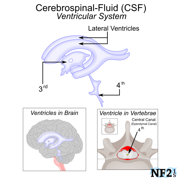
Ventricular System and Cerebrospinal-Fluid Flow Issues
CSF is a clear and colorless fluid that needs to continuously move through a membrane that surrounds the Central Nervous System; brain and spinal cord. Constant CSF movement from the brain to the spinal cord is important for the following:
- Blood Flow: As CSF circulates inside the blood-brain barrier between the brain and spine it helps regulate the consistent flow of blood for nerves, veins, and arteries.
- Giving: CSF helps filter nutrients to the brain.
- Removing: CSF helps filter chemicals and waste matter away from the brain
- Protection: CSF is a protective barrier for the brain and spine that absorbs shock as the body moves or is moved.
Tumor growth can cause issues with the necessary flow of Cerebrospinal Fluid (CSF) from the brain into the spine. There are two forms of Cerebrospinal-Fluid flow issues that might develop; Intracranial Pressure (Obstructive Hydrocephalus) and CSF Leak Intracranial Hypotension).

2. Intracranial Pressure (Obstructive Hydrocephalus)
There are different forms and potential reasons for intracranial pressure (hydrocephalus). For individuals with neurofibromatosis type 2 (NF2), the form of hydrocephalus to develop non-communicating, obstructive hydrocephalus; is an abnormal buildup of CSF in the cavities (ventricles) of the brain. The buildup is often caused by an obstruction of a tumor or multiple tumors which prevents proper fluid drainage. The fluid buildup can raise intracranial pressure inside the skull and result in brain matter compression.
CSF needs to continuously flow from brain to spine and as tumor burden increases, where the flow stops can be fatal. There are warning signs that this is happening and different treatment options.
Signs of Intracranial Pressure (Obstructive Hydrocephalus)
- Papilledema - Optic disc swelling that would be seen by an exam done by a Neuro-Ophthalmologist.
- Severe Headaches these might go away when laying down and return when in an upright position
- Head pressure following; coughing, sneezing, or exertion.
- Tinnitus and Hearing Loss
- Vertigo, Nausea and Vomiting
- Stiff Neck
- Bacterial Meningitis
Possible Treatments for Intracranial Pressure
- Prescription
- Diamox (Acetazolamide), or Lasix (Furosemide) can be used to delay surgical intervention.
- Surgery
- Tumor Removal
- CSF Drain Options
- Lumbar Puncture
- Shunt in Brain MRI Safe type should be used
3. CSF Leak (Intracranial Hypotension/CSF Rhinorrhea)
CSF is a liquid barrier between the brain and skull, as well as a barrier between the spinal cord and vertebrae. During brain and spine surgery, when tumors are removed, the barrier needs to be opened for the operation and closed when completed.
Despite steps taken to close this barrier, if swelling occurs after surgery, the surgery entry point can be easily reopened. This would change the path of normal CSF flow, allowing this vital system fluid to escape.
Intracranial hypotension (CSF Leak), is a result of escape of the fluid from the membrane that surrounds the brain and spinal cord. In addition to closing the entry point with surgical methods, newer additional techniques of brain surgery include using abdominal fat to be added in place of some of the tumor mass removed to prevent this from happening. Most commonly when a CSF Leak occurs it is within 10 days of brain surgery.
Since many of the symptoms of a CSF leak are similar to after surgery issues. In many cases it is not noticed unless bacterial meningitis develops, but can also become evident if there are signs of a sinus issue.
Signs of CSF Leak
- Sinus Drip - Overflow of CSF can force its way through the sinuses.
- Sinus Drip can also leak into the eyes, ear, or nose, but is uncommon.
- Severe Headaches these might go away when laying down and return when in an upright position
- Tinnitus, Balance, and Hearing Problems
- Headache, Nausea, Vertigo, Light Sensitivity, Nausea, and Vomiting
- Bacterial Meningitis
- Visual Disturbances (blurred and or double vision)
- Watery Drainage from the nose (most commonly one side)
- Salty Taste in the Back of the Throat
- Neck Stiffens
Possible Treatments for a CSF Leak
- Natural Method
- Minimal Leakage can heal from bed rest and an increase in fluids.
- Prescription
- Either Diamox (Acetazolamide), or Lasixcan be used to delay surgical intervention. Acetazolamide is a "water pill" (diuretic).
- CSF Drain Options
- Lumbar Puncture
- Shunt in Brain - MRI Safe type should be used
Surgical Methods to Prevent CSF Leak
- Autologous Fat Grafting (free fat graft):
Method used to help fill newly-created dead space, when done so with brain surgery helps
guarantee closure of the dura to prevent CSF Leak.
When done fat is typically used from the individual's abdomen. Found to be a better method than titanium mesh as mesh results in risk of infection. Synthetic options also possible alternatives, but are higher cost.
This can also be used to plug of the bone, or muscular defect created during surgery. Sometimes also used for breast augmentation following breast cancer surgery. (Ackerman, 2013) - Surgical Drains: Placement of drain during surgery to be removed after CSF stops filling the drain
bag 24 or more hours after surgery.
This is a recommended against treatment option unless a brain incision is of considerable size, example of from ear to ear. Other methods would be best used for smaller incisions.
3. Glossary
- cerebrospinal fluid (CSF): a fluid largely secreted by the choroid plexuses of the ventricles of the brain, filling the ventricles and the subarachnoid cavities of the brain and spinal cord. [1]
- cerebrospinal fluid otorrhea: discharge of cerebrospinal fluid through the external auditory canal or through the eustachian tube (ear) into the nasopharynx (back of the nose). [1]
- cerebrospinal fluid rhinorrhea: a discharge of cerebrospinal fluid from the nose. [1]
- blood-cerebrospinal fluid barrier: a barrier located at the tight junctions that surround and connect the cuboidal epithelial cells on the surface of the choroid plexus; capillaries and connective tissue stroma of the choroid do not represent a barrier to protein tracers or dyes. [1]
- meningitis:
Meningitis is inflammation of the membranes of the brain or spinal cord. [1]
A disease marked by inflammation of the meninges (the three membranes covering the brain and spinal cord; the dura, arachnoid, and pia. [3]) that is either a relatively mild illness caused by a virus or severe usually life-threatening illness caused by a bacteria. [2]
Note: Meningitis is often marked by fever, headache, vomiting, malaise, and stiff neck, and if left untreated in bacterial forms, may progress to confusion, stupor, convulsions, coma, and death. [2] - papilledema: swelling and protrusion of the blind spot of the eye caused by edema (abnormal infiltration). [2]
- hydrocephalus: an abnormal increase in the amount of cerebrospinal fluid within the cranial cavity (as from obstructed flow, excess production, or defective absorption) that is accompanied by an expansion of the cerebral ventricles and often increased intracranial pressure, skull enlargement, and cognitive decline [2]
4. Source
-
Dunn, Laurence T. "Raised intracranial pressure." Journal of Neurology, Neurosurgery & Psychiatry 73.suppl 1 (2002): i23-i27.
https://jnnp.bmj.com/content/73/suppl_1/i23 - Medlexicon. "Dictionary." Source: https://www.medilexicon.com/dictionary
- Merriam-Webster. "Medical Dictionary." Source: https://www.merriam-webster.com/medical
- Medical Dictionary. "The Free Dictionary." Source: http://medical-dictionary.thefreedictionary.com
-
Ackerman, Paul D., et al. "The use of abdominal free fat for volumetric augmentation and primary dural closure in supratentorial
skull base surgery: managing the stigma of a temporal defect." Journal of neurological surgery. Part B, Skull base
73.2 (2012): 139.
Source: https://www.ncbi.nlm.nih.gov/pmc/articles/PMC3424629/ | DOI: 10.1055/s-0032-1301399 - U.S. National Library of Medicine. National Institutes of Health. (May 2014). "Normal pressure hydrocephalus (NPH) - Hydrocephalus - idiopathic; Hydrocephalus - adult; Hydrocephalus - communicating; Extraventricular obstructive hydrocephalus."
- U.S. National Library of Medicine. National Institutes of Health. (May 2014). "CSF leak."


 |Google Play